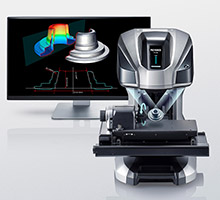
Unlike traditional optical profiling systems that are hard to use and have material restrictions due to low dynamic range, the VR-6000, from Keyence Corporation of America, offers a simple place-and-press interface partnered with HDR scanning to capture highly accurate data, even on glossy or matte surfaces. The HDR algorithm automatically determines optimum lighting conditions, adjusting…
World’s first Microhub makes spatial context accessible for all
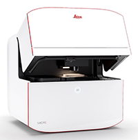
Leica Microsystems, a provider of microscopy and scientific instrumentation and a Life Sciences company of Danaher Corporation, has launched Mica, the world’s first Microhub. A Microhub is a new type of wholly integrated imaging solution that leverages machine learning software, automation tools, and unique fluorescence unmixing techniques to automate the imaging workflow for researchers, regardless…
Thermo Scientific Centrios HX offers precise circuit edit solution for fast prototyping
Thermo Fisher Scientific introduces the Thermo Scientific Centrios HX Circuit Edit System. This state-of-the-art circuit edit solution allows semiconductor manufacturers to optimize success rates with high resolution imaging and precise editing of today’s leading-edge devices. As semiconductor devices become more complex, higher precision circuit edit tools are required to optimize product functionality and deliver prototypes…
R&D 100 of the day: The Neutron and Gamma Ray Source Localization and Mapping Platform 2.0
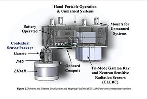
The Neutron and Gamma Localization and Mapping Platform (NG-LAMP), developed by Lawrence Berkeley National Laboratory, is the first ever portable system for simultaneous imaging and mapping of gamma ray and neutron radioactivity in three-dimensions (3D) and in real-time. Unlike all other portable, commercially available radiation imaging systems, which image and map radiation signatures in only…
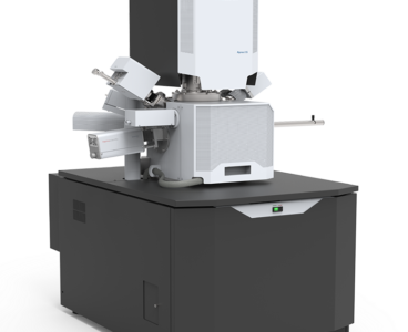
JEOL’s new Scanning Electron Microscope has “Simple SEM” automation and live elemental and 3D Analysis
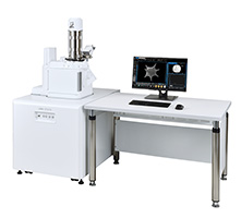
JEOL, developer of cutting-edge electron microscopes for materials characterization and analysis, introduces its latest SEM, the JSM-IT510. This new Scanning Electron Microscope (SEM) features productivity-enhancing automation, including “Simple SEM” automated imaging, automated montaging (both image and EDS map) and live EDS analysis (spectrum and map). The IT510 is the successor to the popular JEOL IT500…
Improved image quality and resolution for all ZEISS laser scanning microscopes
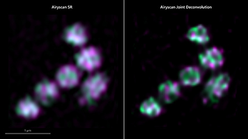
ZEISS has recently introduced two new software functionalities with ZEISS LSM Plus and ZEISS Airyscan Joint Deconvolution, users achieve better results in confocal microscopy. Maximizing recovery requires improvements in efficiency Laser scanning microscopes (LSMs) are the multifunctional high-end microscopes in every imaging facility, valued as much for their instant, high-quality imaging of optical sections as…
The new eviXscan 3D FinePrecision scanner is tiny but has large capabilities
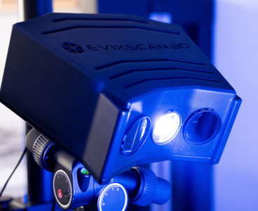
eviXscan 3D FinePrecision scanner, from Evatronix, is an optical measuring device operating with a blue LED light source. The scanner, equipped with two fast, latest generation 8.9 Mpix cameras with CMOS matrices, is characterized by high accuracy reproduction of even the smallest elements of precision mechanics. FinePrecision technology enables an accurate measurement of the dimensions…
Atik Cameras supports the COVID-19 fight with wide range of imaging solutions for use with PCR Instruments
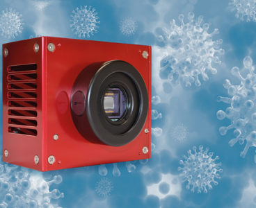
Atik Cameras, a designer and manufacturer of advanced scientific imaging solutions, has further expanded its collaboration with leading global Original Equipment Manufacturers (OEMs) of real time Polymerase Chain Reaction (PCR) DNA amplifiers, securing multiple new contracts to supply high-performing cameras for reliable COVID-19 testing. Under the new agreements, OEMs will access Atik Cameras’ robust range…
Stand-alone microscope camera from Leica Microsystems offers flexibility for imaging tasks
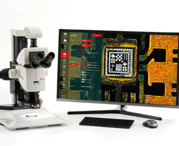
By Maximilian Breuer Product Manager Applied Microscopy, Leica Microsystems There are numerous applications for optical microscopes, ranging from industrial production processes to research and even education. Indeed, they play a vital role in the quality control of final products and components, such as those produced in the electronics industry. Microscopic inspection for quality control enables…
Berkeley Lab team introduces new approach for whole-cell visualization, using the world’s first soft X-ray tomography (SXT) microscope

By Aliyah Kovner The planet is comprised of continents and islands, each with unique cultures and resources. One area may be well known for growing food, another for manufacturing building materials, and yet despite their differences and distance from one another, the regions are linked by global processes. Living cells are built on a similar…
What is hyperspectral image analysis?
An imaging technique that shows the underlying spectrum for each pixel Hyperspectral imaging combines digital imaging with spectroscopy, so that the underlying frequencies in the spectrum for each pixel can be identified. Because only a single wavelength can be represented as a colour for a pixel, a two-dimensional hyperspectral image effectively represents three-dimensional information, in…
R&D 100 winner of the day: VHX-7000 Digital Microscope
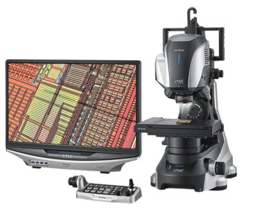
Keyence Corporation of America’s VHX-7000 Digital Microscope pushes the benchmark for what can be offered from a digital microscope. The VHX-7000 is the world’s first 4K microscope, allowing users to view, capture and measure at any angle. With an easy-to-use interface, anyone is able to take high-resolution images and perform 2D/3D measurements. Specialized lenses have…
Engineers produce a fisheye lens that’s completely flat

By Jennifer Chu | MIT News Office To capture panoramic views in a single shot, photographers typically use fisheye lenses — ultra-wide-angle lenses made from multiple pieces of curved glass, which distort incoming light to produce wide, bubble-like images. Their spherical, multipiece design makes fisheye lenses inherently bulky and often costly to produce. Now engineers…
Quantum light squeezes the noise out of microscopy signals
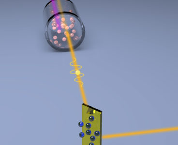
Researchers at the Department of Energy’s Oak Ridge National Laboratory used quantum optics to advance state-of-the-art microscopy and illuminate a path to detecting material properties with greater sensitivity than is possible with traditional tools. “We showed how to use squeezed light – a workhorse of quantum information science – as a practical resource for microscopy,”…
Sensors of world’s largest digital camera snap first 3,200-megapixel images at SLAC
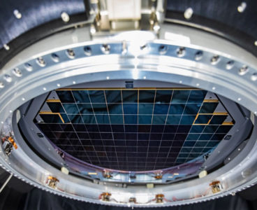
Crews at the Department of Energy’s SLAC National Accelerator Laboratory have taken the first 3,200-megapixel digital photos – the largest ever taken in a single shot – with an extraordinary array of imaging sensors that will become the heart and soul of the future camera of Vera C. Rubin Observatory. The images are so large that…
Super-resolution imaging breakthrough in living cells
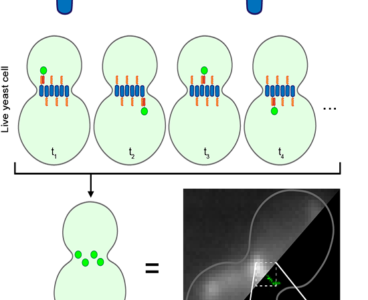
Edinburgh scientists have developed a new imaging technique that reveals the inner workings of living cells in stunning detail and could pave the way to a better understanding of many diseases. The new super-resolution imaging technique – LIVE-PAINT- provides a flexible and powerful way of tracking individual proteins inside living cells, without disrupting their activity.…
New Thermo Scientific Selectris filters push Cryo-EM boundaries with atomic resolution
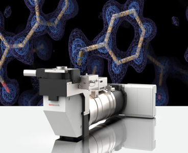
Thermo Fisher Scientific has introduced two breakthrough imaging filters, the Thermo Scientific Selectris Imaging Filter and the Thermo Scientific Selectris X Imaging Filter, taking cryo-electron microscopy (cryo-EM) to a new level, making it possible to view proteins at true atomic resolution. Combined with a Thermo Scientific Krios or Glacios Cryo-Transmission Electron Microscope (TEM), the Selectris…
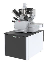
Versatile Thermo Scientific Helios 5 PFIBs enable materials and life sciences research
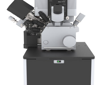
Thermo Fisher Scientific has released the Thermo Scientific Helios 5 Plasma Focused Ion Beam (PFIB) DualBeam and the Thermo Scientific Helios 5 Hydra DualBeam focused ion beam scanning electron microscopes (FIB-SEMs). Built for a wide range of uses, from metals and battery characterization to structural biology, these instruments are well-suited for labs that conduct both materials…
Thermo Fisher Scientific accelerates nanometer-scale research with next-generation Apreo 2
Thermo Fisher Scientific today unveiled its next-generation Thermo Scientific Apreo 2 Scanning Electron Microscope (SEM), a high-performance field emission SEM that takes automation to the next level, offering easy-to-obtain nanometer-scale information at a range of working distances and operating conditions. The Apreo 2 was designed to help investigators, SEM operators and lab managers deliver expert…
JEOL introduces new compact Field Emission Scanning Electron Microscope
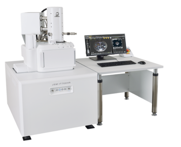
JEOL expands its FE SEM product group with the introduction of a compact Field Emission SEM that offers ultrahigh resolution and versatile analytical capabilities at a great value. The new JSM-IT700HR InTouchScope FE SEM is equipped with a large specimen chamber and both High and Low Vacuum modes for managing a wide variety of specimen…
What is a spectrophotometer?

Spectrophotometers measure a light beam’s intensity as a function of its color (wavelength). Important features of spectrophotometers are spectral bandwidth (the range of colors it can transmit through the test sample), the percentage of sample-transmission, the logarithmic range of sample-absorption, and sometimes a percentage of reflectance measurement. A spectrophotometer is commonly used for the measurement…
What is a spectrometer?
A spectrometer is any instrument used to measure the wavelengths and intensities of a physical characteristic over a given range, i.e. spectrum. The property being measured is usually intensity of light, but other variables like polarization can also be measured. Technically, a spectrometer can function over any range of light, but most operate in a…
Microscopy – Images in a flash
Oak Ridge National Laboratory researchers have built a novel microscope that provides a “chemical lens” for viewing biological systems including cell membranes and biofilms. The tool could advance the understanding of complex biological interactions, such as those between microbes and plants. The noninvasive instrument, detailed in Optics Letters, allows researchers to capture images using ultrashort…
New application for trinamiX near-infrared spectroscopy solutions: plastic sorting made easy

trinamiX, a wholly owned subsidiary of BASF, today announced it has developed a new application for its mobile Near-Infrared (NIR) Spectroscopy Solutions to simplify plastic sorting and recycling. Using trinamiX technology, the diverse compositions of different plastics can now be precisely determined and thus distinguished via the simple use of a portable handheld device that…
JEOL begins remote demos of new ultrahigh resolution FE SEM

JEOL’s new Field Emission Scanning Electron Microscope, the JSM-IT800, is the company’s top-of-the-line microscope with ultrahigh spatial resolution imaging and analysis at the nanoscale. Capabilities include up to 2,000,000 x magnification and an accelerating voltage range of 0.01 to 30 kV, making it possible to acquire stunning details of nanostructures as well as comprehensive analysis…